High Back Muscles Diagram / Iliocostalis Pain Treatment | What Causes It And How To ...
High Back Muscles Diagram / Iliocostalis Pain Treatment | What Causes It And How To .... Muscle of the neck and head 12 photos of the muscle of the neck and head muscle of the head and neck quiz, muscles of the back of neck and head, muscles of the head and neck region, muscles of the head neck and face lateral view, picture muscles of the neck and head, human … The back is the body region between the neck and the gluteal regions. The trapezius and latissimus dorsi muscles connect the upper limb to the vertebral column. On this page, you'll learn about each of these muscles, their locations and functional anatomy. Function of the back muscles there are several individual muscles within the back anatomy, and it's important to take a quick look at all of
ads/bitcoin1.txt
With the back, it's even more difficult… It is opposite from the chest, and the vertebral column runs down the back. Want to maintain muscle flexibility, reduce pain and improve mobility? Shoulder muscle anatomy skeletal muscle anatomy muscular system anatomy human muscle anatomy shoulder muscles leg muscles diagram muscle diagram upper limb anatomy anatomy back. This helps concentrate more stress on the back muscles.
muscles of the arm anterior view - ModernHeal.com from www.modernheal.com Related posts of back muscles chart muscle system diagram. Causes of upper back pain include herniated discs, muscle overuse, osteoarthritis, and a pinched nerve. This muscle diagram made to look like a human. These structures work together to support the body, enable a range of movements, and send messages from the brain to. Both the deltoid and the trapezius are firmly attached to the spine of the scapula. The thoracic spine starts beneath the neck and is comprised of 12 vertebrae, labeled t1 through t12, which go down the back of the torso (figure 1).unlike the cervical spine and lumbar spine, the thoracic spine is relatively immobile because each of its vertebrae are connected to a pair of ribs (one on. These muscles are also called immigrant muscles, since they actually represent true muscles of the back that lie deep to the thoracolumbar fascia. Muscles of the back image search results.
We hope this picture anatomy of back muscles diagram can help you study and research.
ads/bitcoin2.txt
Causes of upper back pain include herniated discs, muscle overuse, osteoarthritis, and a pinched nerve. It is very stiff, and the thoracic spine has a limited range of motion. The human back extends from the buttocks to the posterior portion of the neck and shoulders. The back anatomy includes the latissimus dorsi, trapezius, erector spinae, rhomboid, and the teres major. Nerves in your lower back. The back consists of the spine, spinal cord, muscles, ligaments, and nerves. This diagram shows which muscles in the lower back may be causing you pain. These muscles are also called immigrant muscles, since they actually represent true muscles of the back that lie deep to the thoracolumbar fascia. It is like that for several reasons, all of which you can understand by looking at the anatomy of the thoracic spine. The thoracic spine starts beneath the neck and is comprised of 12 vertebrae, labeled t1 through t12, which go down the back of the torso (figure 1).unlike the cervical spine and lumbar spine, the thoracic spine is relatively immobile because each of its vertebrae are connected to a pair of ribs (one on. The trapezius and latissimus dorsi muscles connect the upper limb to the vertebral column. This helps concentrate more stress on the back muscles. For more anatomy content please follow us and visit our website:
Shoulder muscle anatomy skeletal muscle anatomy muscular system anatomy human muscle anatomy shoulder muscles leg muscles diagram muscle diagram upper limb anatomy anatomy back. If you like training back on machines, try youtube star calum von moger's full program building von moger: These structures work together to support the body, enable a range of movements, and send messages from the brain to. The thoracic spine starts beneath the neck and is comprised of 12 vertebrae, labeled t1 through t12, which go down the back of the torso (figure 1).unlike the cervical spine and lumbar spine, the thoracic spine is relatively immobile because each of its vertebrae are connected to a pair of ribs (one on. The hamstrings are three muscles at the back of the thigh that affect hip and knee movement.
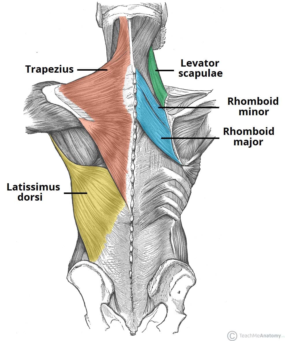
The Superficial Back Muscles - Attachments - Actions ... from teachmeanatomy.info Related posts of back muscles chart muscle system diagram. Another common cause of lower back and hip pain is disc injury. The pelvis at the bottom of the back and the shoulders at the top of the back give the back. Shoulder muscle anatomy skeletal muscle anatomy muscular system anatomy human muscle anatomy shoulder muscles leg muscles diagram muscle diagram upper limb anatomy anatomy back. This helps concentrate more stress on the back muscles. It's sometimes hard to explain the pain to someone else, because it only occurs during certain activities, or at some point in the normal range of motion of the muscle or muscle group. By the way, have you heard about the myth of. The extrinsic back muscles, which lie most superficially on the back.
Keep your chest out and flexed throughout the move;
ads/bitcoin2.txt
Moves humerus (arm) to chest. Causes of upper back pain include herniated discs, muscle overuse, osteoarthritis, and a pinched nerve. The back is the body region between the neck and the gluteal regions. Muscle system diagram 12 photos of the muscle system diagram muscular system diagram blank, muscular system diagram front and back, muscular system diagram printable, muscular system key diagram 2, printable muscular system diagram download, human muscles, muscular system diagram blank, muscular system diagram front and back. It comprises the vertebral column (spine) and two compartments of back muscles; Anatomynote.com found anatomy of back muscles diagram from plenty of anatomical pictures on the internet. See back muscles and low back pain. Both the deltoid and the trapezius are firmly attached to the spine of the scapula. The extrinsic back muscles, which lie most superficially on the back. If you like training back on machines, try youtube star calum von moger's full program building von moger: Function of the back muscles there are several individual muscles within the back anatomy, and it's important to take a quick look at all of Middle back pain can be caused by strain from daily activities and poor posture, a past or recent injury, or muscle inflammation. Related posts of back muscles chart muscle system diagram.
Read below for more information on why you may be having prolonged or sudden pain in the middle of your back, related symptoms, and treatment options. In this image, you will find frontalis, orbicularis oculi, zygomaticus, masseter, orbicularis oris, sternocleidomasteoid, deltoid, pectoralis major, biceps brachii, iliopsoas, adductor longus, gastrocnemius. Anatomynote.com found anatomy of back muscles diagram from plenty of anatomical pictures on the internet. Quadriceps (made of 4 muscles): Five pairs of lumbar spinal nerves labeled l1 to l5 branch off your spinal cord and exit through small holes between the vertebrae.
Pin by ashlee brown on nursing :) | Leg muscles anatomy ... from i.pinimg.com The back anatomy includes the latissimus dorsi, trapezius, erector spinae, rhomboid, and the teres major. Daniel nelson on january 1, 2019 2 comments 🔥! This muscle diagram made to look like a human. These muscles are also called immigrant muscles, since they actually represent true muscles of the back that lie deep to the thoracolumbar fascia. Quadriceps (made of 4 muscles): We think this is the most useful anatomy picture that you need. The hamstrings are three muscles at the back of the thigh that affect hip and knee movement. The human back extends from the buttocks to the posterior portion of the neck and shoulders.
The hamstrings are three muscles at the back of the thigh that affect hip and knee movement.
ads/bitcoin2.txt
Having mid back pain is a common condition that can also feel like tightness or tension in the center of your back. For more anatomy content please follow us and visit our website: Most of the time, back muscle pain is diagnosed then treated with little more than a prescription of rest, painkillers and muscle relaxants. This diagram shows which muscles in the lower back may be causing you pain. The extrinsic back muscles, which lie most superficially on the back. Muscle system diagram 12 photos of the muscle system diagram muscular system diagram blank, muscular system diagram front and back, muscular system diagram printable, muscular system key diagram 2, printable muscular system diagram download, human muscles, muscular system diagram blank, muscular system diagram front and back. The thoracic spine starts beneath the neck and is comprised of 12 vertebrae, labeled t1 through t12, which go down the back of the torso (figure 1).unlike the cervical spine and lumbar spine, the thoracic spine is relatively immobile because each of its vertebrae are connected to a pair of ribs (one on. Middle back pain can be caused by strain from daily activities and poor posture, a past or recent injury, or muscle inflammation. See back muscles and low back pain. Moves humerus (arm) to chest. This helps concentrate more stress on the back muscles. Another common cause of lower back and hip pain is disc injury. Both the deltoid and the trapezius are firmly attached to the spine of the scapula.
ads/bitcoin3.txt
ads/bitcoin4.txt
ads/bitcoin5.txt
0 Response to "High Back Muscles Diagram / Iliocostalis Pain Treatment | What Causes It And How To ..."


0 Response to "High Back Muscles Diagram / Iliocostalis Pain Treatment | What Causes It And How To ..."
Posting Komentar